Journal of NeuroEngineering and Rehabilitation provides a forum for researchers and clinicians interested in understanding the way neuroscience and biomedical engineering are continuing to reshape physical medicine and rehabilitation and human movement augmentation. JNER hosts the introduction of new methods and the discussion of their clinical implications, and offers an opportunity to publish, in a timely manner, articles relevant to the intersection of these three fields.
Aims and scope
Vacancy: Editor-in-Chief, Journal of NeuroEngineering and Rehabilitation
BioMed Central is seeking to appoint an Editor-in-Chief to succeed Professor David J Reinkensmeyer, who will be stepping down after a term of 10 years at the helm of JNER.
Find out more about the role and application process.
Article collections
Click here to see the article collections and thematic series that the journal has published in recent years.
Editor's picks
See what recently published articles the Editor in Chief is excited about!
Most cited article published in 2023
Jesús de Miguel-Fernández, Joan Lobo-Prat, Erik Prinsen, Josep M. Font-Llagunes & Laura Marchal-Crespo
Top in Rehabilitation
Journal of NeuroEngineering and Rehabilitation is ranked second in the Rehabilitation category by Scopus!
Articles
-
-
Fatigue in children using motor imagery and P300 brain-computer interfaces
-
Evaluation of power wheelchair driving performance in simulator compared to driving in real-life situations: the SIMADAPT (simulator ADAPT) project—a pilot study
-
A comprehensive multivariate analysis of the center of pressure during quiet standing in patients with Parkinson's disease
-
Use of functional magnetic resonance imaging to identify cortical loci for lower limb movements and their efficacy for individuals after stroke
-
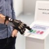
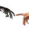
A review of wearable sensors and systems with application in rehabilitation
-
Rehabilitation of gait after stroke: a review towards a top-down approach
-
A wireless body area network of intelligent motion sensors for computer assisted physical rehabilitation
-
The feasibility of a brain-computer interface functional electrical stimulation system for the restoration of overground walking after paraplegia
-
Motor rehabilitation using virtual reality
In Review
Journal of NeuroEngineering and Rehabilitation has launched In Review, a new option that provides authors with on-demand information on the status of their manuscript, enables them to share their work with funders and their research community, and allows their colleagues to comment and collaborate - all whilst their manuscript is under review.
Read moreStandards of reporting
Journal of NeuroEngineering and Rehabilitation advocates the complete and transparent reporting of research and methods. Authors are required to follow relevant reporting guidelines and append the appropriate reporting guideline checklist to their manuscript on submission, available from the EQUATOR Network. See BMC’s policy page for further information.
Person-first language
Journal of NeuroEngineering and Rehabilitation recommends the use of person-first language to speak appropriately about individuals with a disability. For example, when referring to a person with a stroke, refer to the person first using a phrase such as 'a person with a stroke' or 'a person who has a stroke'. Avoid terms such as 'victim', 'patient', 'the handicapped', 'the disabled', 'suffering', or 'brain damaged'.
Authors may wish to refer to the ADA guidelines for writing about people with disabilities.
From the blog
David Reinkensmeyer, Editor-in-Chief
David Reinkensmeyer is a Professor in the Department of Mechanical and Aerospace Engineering, the Department of Anatomy and Neurobiology, and the Department of Biomedical Engineering at University of California, Irvine. Prior to this, he received a B.S. degree in electrical engineering from the Massachusetts Institute of Technology, and M.S. and Ph.D. degrees in electrical engineering from the University of California, Berkeley. He was a postdoctoral fellow then research assistant professor in the Sensory Motor Performance Program, Rehabilitation Institute of Chicago and Department of Physical Medicine and Rehabilitation, Northwestern University Medical School, before joining U.C Irvine in 1998. Prof. Reinkensmeyer's research interests include neuromuscular control, motor learning, robotics, and rehabilitation.
In 2019, David Reinkensmeyer was elected as fellow of American Institute for Medical and Biological Engineering (AIMBE) to recognize his innovative contributions to rehabilitation robotics and neural engineering, and for design of practical devices to enhance patient recovery.
Annual Journal Metrics
-
2022 Citation Impact
5.1 - 2-year Impact Factor
5.8 - 5-year Impact Factor
1.924 - SNIP (Source Normalized Impact per Paper)
1.134 - SJR (SCImago Journal Rank)2023 Speed
19 days submission to first editorial decision for all manuscripts (Median)
225 days submission to accept (Median)2023 Usage
1,800,927 downloads
756 Altmetric mentions